
Nail Anatomy
Nails are important structures that protect the mechanical integrity of the dorsal surfaces of our digits/fingers!
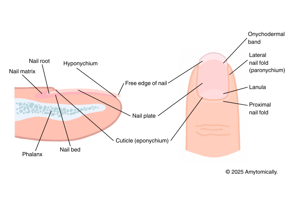
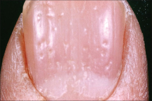
Anatomical landmarks (Figure 1)
Nail plate/body: visible nail portion covering nail bed
Free edge of nail: nail plate hanging past hyponychium
Cuticle (eponychium): thin stratum corneum part of nail root that extends over nail bed
Hyponychium: thickened stratum corneum at tip of finger
Lanula: pale crescent shaped area at bottom of nail (due to obstruction of blood vessels)
Nail folds (lateral, proximal): soft tissue that protects nail edges
Nail root, matrix (discussed below)
Nail production
Nail matrix:
- Responsible for formation of hard nail plate and the production of onychocytes (nail cells) via keratinization which pushes cells further over nail plate distally
- Contains melanocytes
Nail root:
- Deepest part of nail where growth occurs
Pathological Indications
Nail appearance and morphology can often reveal important insights pertaining to infection, systemic conditions, & more. Below is a brief list of some of the most common nail conditions.
Localized conditions
- Melanonychia: dark pigment under nail plate or Hutchinson’s sign (involvement of proximal nail fold) may indicate melanoma
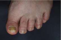
- Onychomycosis: yellow discoloration of nail may indicate fungal infection
- Leukonychia: white discoloration of nail due to abnormal keratinization
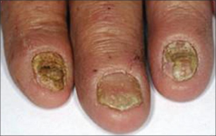
- Paronychia: nail inflammation presenting with crusting; most commonly due to staph. aureus infection
Systemic conditions
- Kolionychia (concave nails): associated with blood disorders
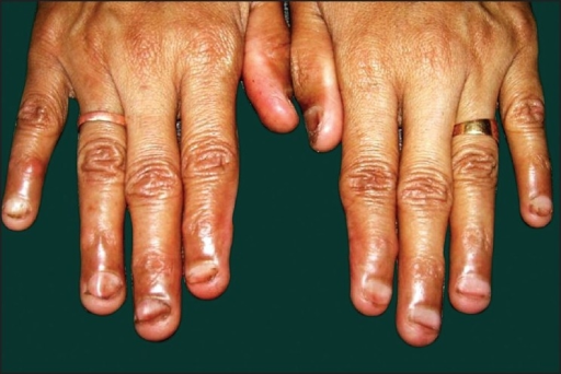
- Clubbing (convex nails): COPD, cirrhosis/liver disease
- Yellow discoloration: chronic respiratory disorders, thyroid disease, AIDs
- Flaking: abnormally low levels of vit A, B12, linoleic acid
- Pitting (nail depressions): psoriasis, alopecia
Nail pathology images

Leave a comment