
The EKG
The electrocardiogram (ECG or EGK) is a non-invasive method of testing the electrical activity of the heart. Notable EKG scans and patterns can reveal incidence of certain disorders.
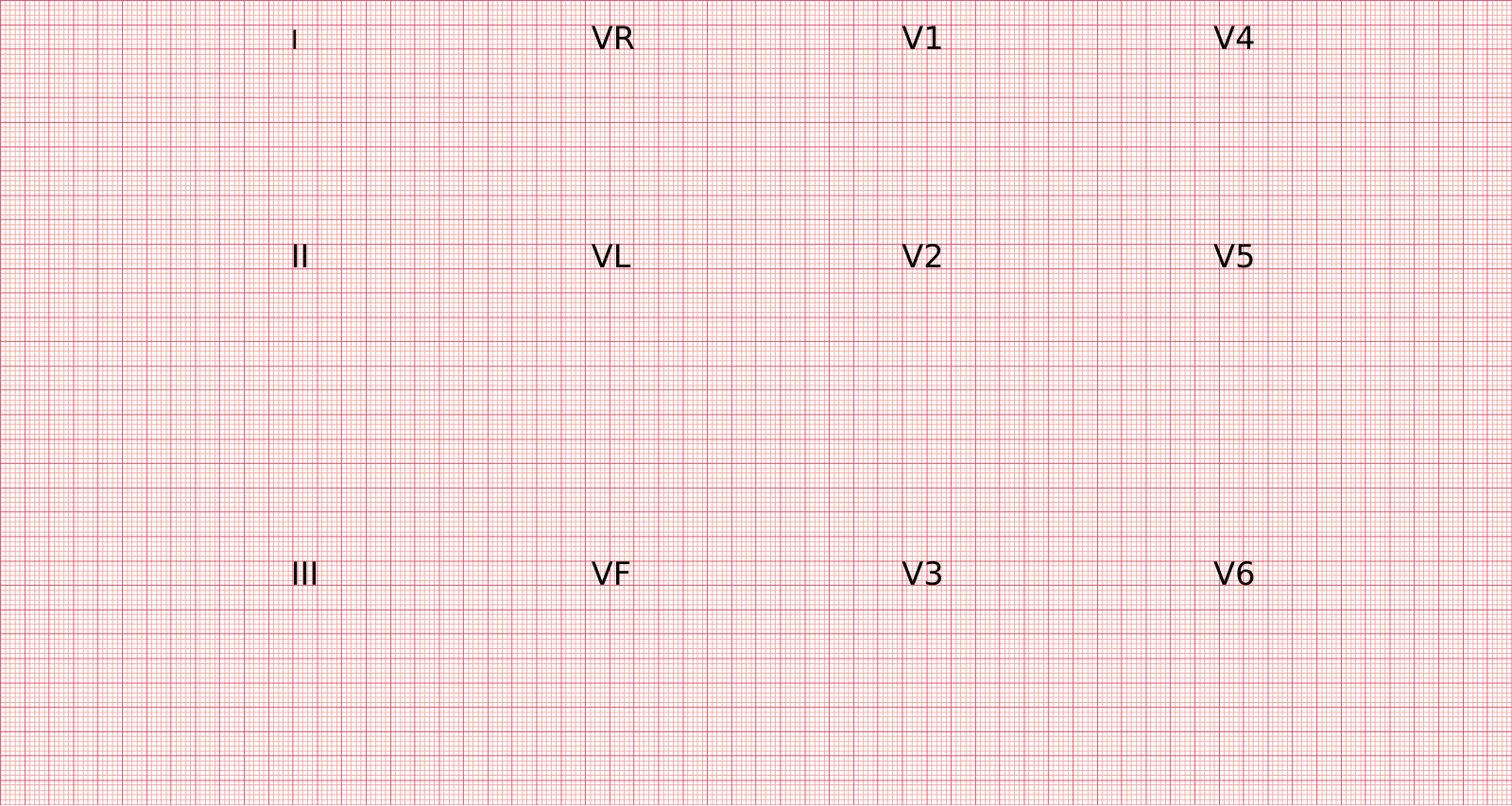
It typically involves the usage of twelve leads:
- Bipolar limb leads (3): I, II, III
- Augmented unipolar limb leads (3): aVF, aVL, aVR
- Precordial chest leads (6): V1, V2, V3, V4, V5, V6
Lead association with heart landmarks:
- Inferior wall: leads II, III, aVF
- Lateral wall: I, aVL, V5-6
- Right ventricle: primarily V1
- Anteroseptal: V1-V4

Waves
The isoelectric line is the baseline for which waves fluctuate off of (i.e., the sole line present in the absence of heart electrical activity).
P Wave: atrial depolarization
- Upright in leads I, II and inverted in aVR
QRS Complex: ventricular depolarization
- Normal duration: 0.06-0.10s
- Wide: >0.12s; Narrow: <0.12s
- Q wave
- Indicate possible pathology if seen in V1-3
- Low voltage QRS
- Summation of R waves in leads I, II, III is <15mm
- Indicate pericardial effusion, amyloidosis or sarcoidosis, COPD, CHF
T Wave: ventricular repolarization
- Upright in most leads, and inverted in aVR
- Inversion in aVL can indicate inferior wall MI
- Hyperacute (broad, tall, asymmetrical): can indicate early STEMI, vasospasm
- Flat: impending ischemia, hyperkalemia
Segments vs. Intervals
Segments indicate the region between two waves. Intervals encompass the waves as well as the segment in between.
Segments
PR segment:
- Typically flat
- Depression can indicate pericarditis
ST segment:
- J point: begins at ST; can be used as metric for ST depression or elevation
- ST depression: If the J point is >0.5mm below isoelectric in two continuous leads
- ST elevation: J point >1mm above isoelectric lines in leads except V2, V3
Intervals
RR interval:
- The distance between two R waves (and thus is a measure of the entire cycle)
- Determines rhythm regularity
- A constant RR interval indicates regular rhythm; a non-constant interval indicates irregularity
PR interval:
- <0.2s under normal conditions
- Can help indicate atrial enlargement
- Right atrial enlargement (RAE): lead II P wave is ≥2.5mm above isoelectric line
- Left atrial enlargement (LAE): bifid P wave in II
QT interval:
- Typically is half of RR interval
- Prolonged QT interval: >460ms (females), >450ms (males); can indicate torsades de pointes (TdP)
- Short: <350ms
Understanding the EKG Strip
The typical EKG rhythm strip presents with 5×5 graph boxes (Figure 2), and they are typically 10 seconds (50 large boxes) long. The horizontal axis measures time in seconds (s), and the vertical axis measures voltage, in millivolts (mV). For the horizontal axis, one small box is the equivalent duration of 0.04s, and thus five small boxes (one large box) equivocates to 0.20s. Voltage, on the vertical axis, is measured with one small corresponding to 0.1mV, and five small boxes (one large box) measuring 0.5mV.
A typical rhythm strip will have an extended and longer strip at the bottom, which is typically lead II (undepicted in Figure 2).
Calculating heart rate (HR): normal HR is typically 60-100 beats per minute (BPM)
- First method: multiply the number of R waves in a 6-second segment of the rhythm strip by 6
- Ex. If there are 10 R waves: 10 x 6 = 60 BPM
- Second method: divide 300 by the number of large boxes in between two R waves
- Ex. If there are 6 large boxes: 300/6 = 50 BPM

Leave a comment