
Heart Valves
Valvular anatomy and contractile mechanism
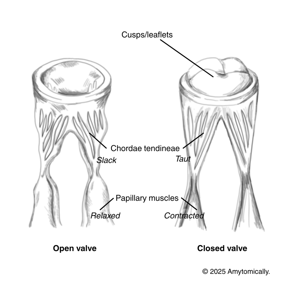
The valves in our hearts are formed of the cusps/leaflets themselves, and are controlled by chordae tendineae and the attached papillary muscles.
- Valvular cusps are fibrous flaps that regulate the passage of blood from one chamber to another, and prevent backflow.
- Chordae tendineae are connective collagenous fibers that anchor the free edges of cusps to prevent back flow (by maintaining tension).
- Papillary muscles are conical muscular projections which insert in the right and left ventricles and contract to close the valve, and relax to open it. They are modified derivatives of the trabeculae carneae, which are the muscular ridges of the interior ventricles.
- In fact,the trabeculae carneae are responsible for carrying the electrical impulse to stimulate contraction of papillary muscles (via the moderator band).
Cusp variation
AV Valves
Tricuspid valve (regulates blood flow from right atrium to ventricle)
- 3 cusps: anterior, posterior, septal cusps
Bicuspid/Mitral valve (from left atrium to ventricle)
- 2 cusps: anterior, posterior cusps
Semilunar Valves
Pulmonary valve (from right ventricle to pulmonary arteries)
- 3 cusps: anterior, left, right semilunar cusps
Aortic valve (from left ventricle to ascending aorta)
- 3 cusps: right coronary, left coronary, non-coronary cusps
- In the disorder bicuspid aortic valve an individual may present with only two cusps due to the fusing of 2

Leave a reply to Aortic Dissection – Amytomically Cancel reply