
Cartilage
Cartilage is found between connective tissue (CT) and bone, and is classified into hyaline, fibrocartilage, and elastic cartilage (discussed in depth below).
Cartilage composition
- Matrix: water, chondroitin sulfate, hyaluronic acid, collagen fibers
- Vasculature: is not vascularized and receives no innervation (thus only receives nutrient via perichondrial diffusion)
Perichondrium: external protective and supportive layer
- Outer layer: fibrous, dense irregular CT (many fibroblasts, T1 collagen)
- Inner layer: chondrogenic layer with chondroblasts and progenitors
- Present around hyaline and elastic cartilage, only
Cartilage growth
Cartilage grows via interstitial (longitudinal growth) and appositional growth (lateral/horizontal). These types of growth are analagous to bone growth at epiphyseal plates during development, and they only occur during development (i.e., under normal conditions, neither interstitial or appositional growth once adulthood is reached)
Interstitial growth: chondrocytes divide, producing matrix
Appositional growth: progenitor cells within inner perichondrial layer differentiate into chondroblasts, which secrete matrix adding onto to width of lateral surfaces
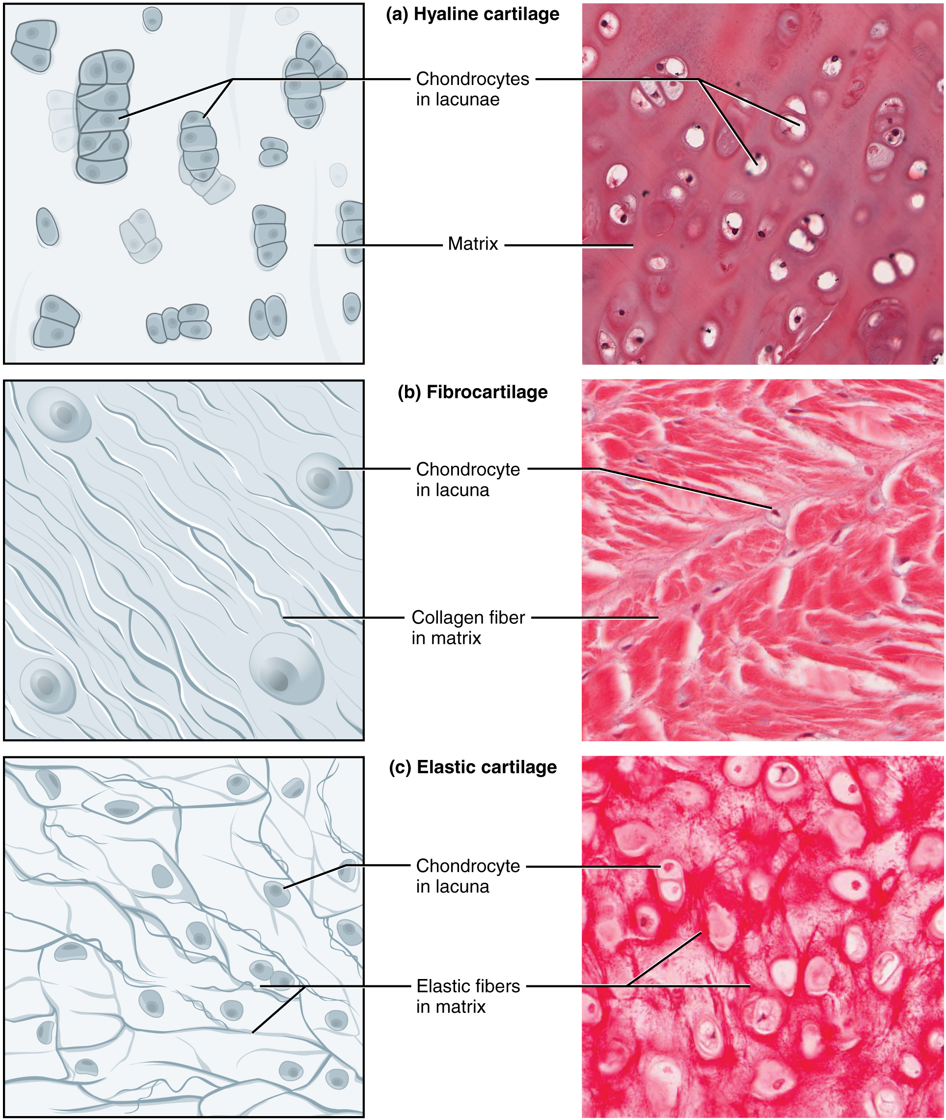
Cartilage Types & Histological Pearls
Hyaline cartilage
- The most abundant type
- Composition: type II (T2) collagen with proteoglycans; few chondrocytes
- Histology: glassy matrix (typically blue/white on gross view) with non-visible collagen fibers
- Functions: minimizes surface friction, so good at resisting compressive forces (e.g., the ends of long bones have hyaline articulate cartilage to absorb compression)
- Locations: embryonic skeleton before ossification, respiratory passages (larynx, trachea), nose bridge, costal cartilage
Fibrocartilage
- Composition: T1 collagen, very little ground substance
- No perichondrium
- Histology: very dense collagen fiber network
- Functions: high compressive/tensile resistance
- Locations: intervertebral discs, knee meniscus, tendons, ligaments
Elastic cartilage
- Composition: T2 collagen, many chondrocytes, more ground substance (with proteoglycans)
- Histology: highly visible elastic fibers
- Functions: strength and flexibility (typically yellow on gross)
- Locations: epiglottis, larynx, eustachian tube, auricle of ear

Leave a comment