
Impetigo
Info & Pathophysiology
What is it?
Highly contagious, purulent, superficial bacterial (G+) infection of the stratum corneum
Risk Factors: young age (increases incidence of bullous form in specific), crowding (due to propagated nature of spread); poor hygiene, warm/humid environment (both increase likelihood of harboring bacteria); burns, trauma, bites
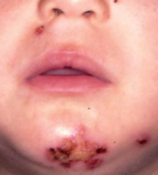
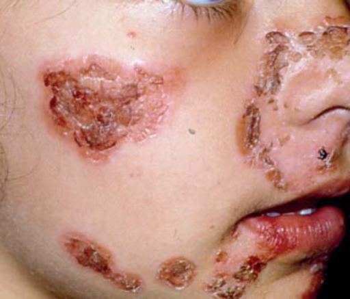
Clinical Diagnosis: typically honey crusted (plaque-like) lesions surrounded by erythematous base
Pathophysiology:
- Primary infectious agents: Staphylococcus aureus , Group A beta-hemolytic Streptococcus/GABHS
- GABHS accesses fibronectin receptors through injury and can self-inoculate
- Primary infection: direct bacterial invasion
- Secondary infection: infection at previous wound site
- Zosteriform impetigo: varicella-zoster virus infection (i.e. shingles) increases susceptibility for developing impetigo; the infection itself will thus also align with dermatomes
Major Subtypes
Nonbullous (70%)
Most common subtype, caused by Group A Strep (GABHS)
Papules progress into vesicles, which coalesce and rupture, forming crust with erythematous base
Lesions on face, extremities, no fever present
Bullous (30%)
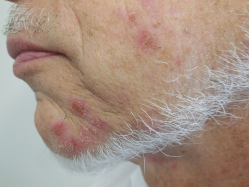
Exclusively caused by an exfoliative toxin of s. aureus
Small vesicles that become flaccid bullae; no honey-colored crust or erythema and fever
Ecthyma (<10%)
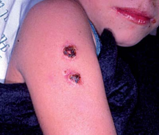
Ulcerative lesions with black holes
Extends deeper (i.e. past epidermis)
More painful than other two types
Treatment
If limited:
- Remove crusts via saline compresses, antiseptic soaks
- Topical treatments: retapamulin 5d, mupirocin 5d, fusidic acid
If extensive or ecthyma presentation:
- Cloxacillin 7-10d
For bullous impetigo or nonbullous impetigo (>5 lesions): treat with systemic antibiotics
- Betalactams: cephalosporins, amoxicillin, dicloxacillin
- MRSA antibiotics

Leave a comment